Bones In Your Leg Diagram / Lower Extremity Anatomy Bones Muscles Nerves Vessels Kenhub / This can cause tenderness in the right or left buttock and severe pain down the leg.
byAdmin•
0
Bones In Your Leg Diagram / Lower Extremity Anatomy Bones Muscles Nerves Vessels Kenhub / This can cause tenderness in the right or left buttock and severe pain down the leg.. Let's review all of these bones one last time. Die heißesten sneaker releases online. The lower limb contains 30 bones. As these muscles contract and relax, they move skeletal bones to create movement of the body. The bones together make up the hip.
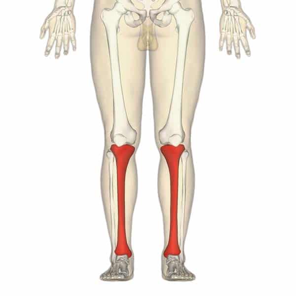
The talus the weight of your body is transferred from the tiba to the talus. Many muscles that move the trunk and legs, such as our abdominal muscles, attach to the hip bones. The foot begins at the lower end of the tibia and fibula, the two bones of the lower leg. The tibia, commonly known as the 'shin bone', is the largest and most medial of the two.you can palpate its anterior border when you run your finger down the anterior aspect of your leg. The hip itself is a ball and socket joint, much like the shoulder.the structures necessary to create this joint are the socket, the joint capsule, muscle, ligaments, and the neck.
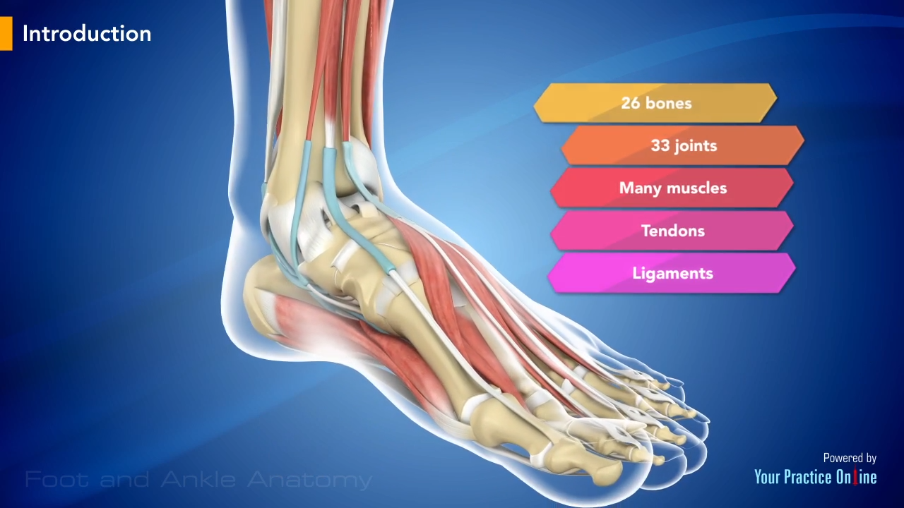
Foot And Ankle Anatomy Video Medical Video Library from www.ypo.education 15 photos of the leg bones anatomy diagram. The bones of the foot are organized into the tarsal bones, metatarsal bones, and phalanges. This allows weight to be distributed either anteriorly or posteriorly throughout the foot. At the same time, the bones and joints of the leg and foot must be strong enough to support the body's weight while remaining. The smaller bone that runs alongside the tibia (fibula) and the kneecap (patella) are the other bones that make the knee joint. The medial, larger bone of the lower leg. Almost every skeletal muscle works by pulling two or more bones either closer together or further apart. The lower extremity, commonly referred to as the leg, contains four bones (the femur, the patella, the tibia, and the fibula) and bends at the hip, the knee, and the ankle.
The bones of the leg and foot form part of the appendicular skeleton that supports the many muscles of the lower limbs.
The lower limb contains 30 bones. The hip itself is a ball and socket joint, much like the shoulder.the structures necessary to create this joint are the socket, the joint capsule, muscle, ligaments, and the neck. The smaller lateral bone of the lower leg. The human leg, in the general word sense, is the entire lower limb of the human body, including the foot, thigh and even the hip or gluteal region. The proximal portion of the tibia is tibial plateau which acts as a cusp for the knee, the distal portion tapers into the medial malleoli and the concave surface which articulates with the talus at the ankle joint. These muscles work together to produce movements such as standing, walking, running, and jumping. The bones of your leg and foot helped give you the ability to score that field goal. The bones of the skeletal system act as attachment points for the skeletal muscles of the body. The talocrual joint is made up of three main bones. That puts pressure on the nerves in the area and can cause pain, tingling, numbness, or weakness in your legs. The foot begins at the lower end of the tibia and fibula, the two bones of the lower leg. Many muscles that move the trunk and legs, such as our abdominal muscles, attach to the hip bones. Your hamstring is directly connected to your ischium bones, and any tear or damage to your hamstring can result in sit bone pain.
Die heißesten sneaker releases online. Your hamstring is directly connected to your ischium bones, and any tear or damage to your hamstring can result in sit bone pain. The bones of the appendicular skeleton provide support and flexibility at the joints and anchor the muscles that move the limbs. Let's review all of these bones one last time. These muscles work together to produce movements such as standing, walking, running, and jumping.
The Tibia Proximal Shaft Distal Teachmeanatomy from teachmeanatomy.info He leg's main function in the human is for locomotion and support of the rest of the body. The human leg consists of 8 bones, 4 per leg. The femur is the single bone of the thigh. The proximal portion of the tibia is tibial plateau which acts as a cusp for the knee, the distal portion tapers into the medial malleoli and the concave surface which articulates with the talus at the ankle joint. The foot begins at the lower end of the tibia and fibula, the two bones of the lower leg. This area is commonly referred to as the calf. The pubis, ischium, and ilium together constitute the pelvis while the thigh bone is the femur. The bones together make up the hip.
Your hamstring is directly connected to your ischium bones, and any tear or damage to your hamstring can result in sit bone pain.
Also called the shin bone, the tibia is the longer of the two bones in the. Die heißesten sneaker releases online. These are the femur, patella, tibia, fibula, tarsal bones, metatarsal bones, and phalanges (see figure 6.51). The bones of the leg are the femur, tibia, fibula and patella.the foot bones shown in this diagram are the talus, navicular, cuneiform, cuboid, metatarsals and calcaneus. The hip itself is a ball and socket joint, much like the shoulder.the structures necessary to create this joint are the socket, the joint capsule, muscle, ligaments, and the neck. The thigh bone, or femur, is the large upper leg bone that connects the lower leg bones (knee joint) to the pelvic bone (hip joint). The bones together make up the hip. Many muscles that move the trunk and legs, such as our abdominal muscles, attach to the hip bones. Let's review all of these bones one last time. The human leg, in the general word sense, is the entire lower limb of the human body, including the foot, thigh and even the hip or gluteal region. The tibia, commonly known as the 'shin bone', is the largest and most medial of the two.you can palpate its anterior border when you run your finger down the anterior aspect of your leg. At the same time, the bones and joints of the leg and foot must be strong enough to support the body's weight while remaining. 15 photos of the leg bones anatomy diagram.
The majority of muscles in the leg are considered long muscles, in that they stretch great distances. The bones of the appendicular skeleton provide support and flexibility at the joints and anchor the muscles that move the limbs. As these muscles contract and relax, they move skeletal bones to create movement of the body. This keeps the bones together, giving a high ankle sprain time to heal. The femur is the single bone of the thigh.
The Knee Anatomy Injuries Treatment And Rehabilitation from i0.wp.com The lower leg contains two major long bones, the tibia and the fibula, which are both very strong skeletal structures. The knee joint is the largest joint in the body and is primarily a hinge joint, although some sliding and rotation occur. Browse 7,053 leg bone stock photos and images available, or search for human leg bone or leg bone xray to find more great stock photos and pictures. Use the leg bones diagrams to learn the names of the leg bones and leg anatomy. The tibia, commonly known as the 'shin bone', is the largest and most medial of the two.you can palpate its anterior border when you run your finger down the anterior aspect of your leg. The sacrum and the coccyx attach to the two hip bones to form the pelvis, but are more important to the spinal column, where they are counted. Distal to the ankle is the foot. 15 photos of the leg bones anatomy diagram.
The bones of the leg and foot form part of the appendicular skeleton that supports the many muscles of the lower limbs.
Femur (2 bones) patella or kneecap (2 bones) tibia (2 bones) fibula (2 bones) foot (52 bones in total, 26 per foot) tarsus/tarsals. Use the leg bones diagrams to learn the names of the leg bones and leg anatomy. The lower leg extends from the knee to the ankle. Diagram and names of leg bones, diagram of foot and leg bones, diagram of leg bones, diagram of lower leg bones, diagram of the bones in your … source: The proximal portion of the tibia is tibial plateau which acts as a cusp for the knee, the distal portion tapers into the medial malleoli and the concave surface which articulates with the talus at the ankle joint. The bones of the hip include the femur, the ilium, the ischium, and the pubis. Your hamstring is directly connected to your ischium bones, and any tear or damage to your hamstring can result in sit bone pain. Related posts of diagram of leg bones bone anatomy elbow. The bones of the leg and foot form part of the appendicular skeleton that supports the many muscles of the lower limbs. Browse 7,053 leg bone stock photos and images available, or search for human leg bone or leg bone xray to find more great stock photos and pictures. This keeps the bones together, giving a high ankle sprain time to heal. The talus the weight of your body is transferred from the tiba to the talus. Ankle & lower leg anatomy.
The lower leg extends from the knee to the ankle bones in leg diagram. The bones of the leg and foot form part of the appendicular skeleton that supports the many muscles of the lower limbs.